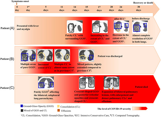
Figure 2.

The CT scan findings of COVID‐19 progression from three patients. Patient (A) was a 61‐year‐old male with a history of chronic pleurisy. CT also shows calcified fibrothorax in the left lob due to chronic pleurisy. 63 Patient (B) was a 35‐year‐old female. 64 Patient (C) was a 77‐year‐old male with cerebrovascular disease, cardiovascular disease, and hypertension. 49 CL, consolidation; CT, computed tomography; GGO, ground‐glass opacity; PNA, pneumonia [Color figure can be viewed at wileyonlinelibrary.com]
