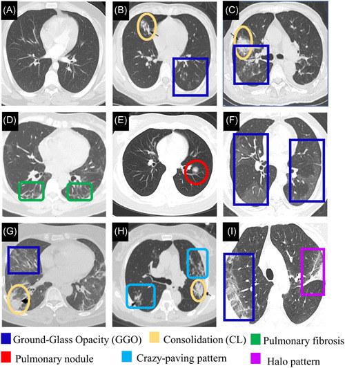
Figure 3.

(A) CT image of normal sample. 19 CT images from pneumonia patients (Adapted from Reference [66]. (B) GGO and consolidation are shown in a 23‐year‐old female with influenza infection. (C) a mixed pattern of consolidation and GGO is shown in a 64‐year‐old male with Epstein‐Barr virus infection. CT images from confirmed COVID‐19 patients. (D) Pulmonary fibrosis is shown in both lungs in a CT image of a 56‐year‐old female with moderate COVID‐19 (Adapted from Reference [62]). (E) Pulmonary nodule in a 23‐year‐old female (Adapted from Reference [67]). (F) Confluent pure GGO is presented in COVID‐19 patient (Adapted from Reference [68]). (G) GGO and consolidation with air bronchogram are presented in a CT image of 59‐year‐old female with sever COVID‐19 (adapted from 62 ). (H) Multifocal crazy‐paving pattern and consolidation is shown in a 59‐year‐old female (Adapted from Reference [66]). (I) the right lung with peripheral predominant GGO, and a reversed halo sign in the left in a 45‐year‐old female (Adapted from Reference [69]). CL, consolidation; CT, computed tomography; GGO, ground‐glass opacity [Color figure can be viewed at wileyonlinelibrary.com]
