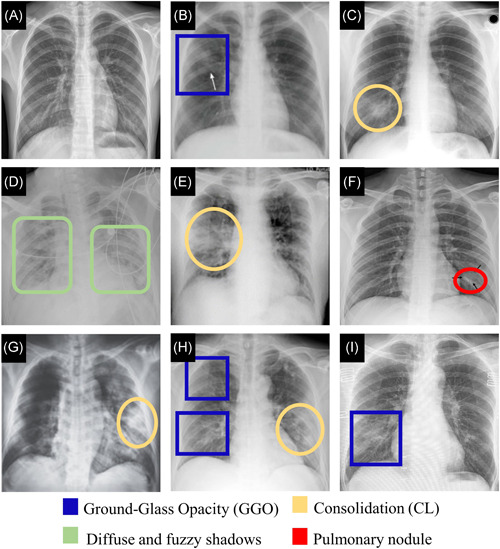
Figure 4.

Chest X‐ray images. (A) Normal sample. 92 (B) A female in her 30 s with Mycoplasma pneumoniae pneumonia. X‐ray shows GGO (Adapted from Reference [93]). (C) A 29‐year‐old female with SARS. X‐ray reveals consolidation (Adapted from Reference [94]). (D) A 41‐year‐old male with COVID‐19. X‐ray, obtained 11 days after the symptoms, shows bilateral diffuse patchy and fuzzy shadows (Adapted from Reference [95]). (E) An elderly male with COVID‐19. X‐ray shows consolidative changes in the right lobe (Adapted from Reference [91]). (F) A single nodular consolidation is observed in X‐ray image from a patient with COVID‐19 (Adapted from Reference [68]). (G) A 65‐year‐old male. X‐ray, obtained 9 days after onset of symptoms, shows a progressive infiltrate and consolidation (Adapted from Refernce [96]). (H) X‐ray of patient of COVID‐19 shows patchy ground‐glass opacities and patchy consolidation (Adapted from Reference [68]). (I) A 59‐year‐old female with COVID‐19. X‐ray shows patchy GGO (Adapted from Reference [69]). CL, consolidation; CT, computed tomography; GGO, ground‐glass opacity [Color figure can be viewed at wileyonlinelibrary.com]
