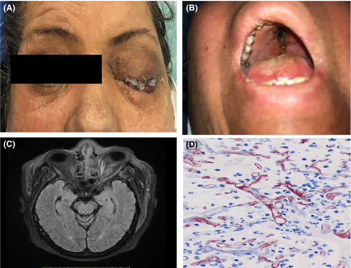
FIGURE 1.

Clinical, radiological and histological features in one of our patients with COVID‐19‐associated mucormycosis. (a) Complete eyelid ptosis, restricted eye movements and no‐light perception in left eye. (b) Palate eschar. (c) Brain MRI, T1‐weighted image after gadolinium injection revealed left ethmoid sinus opacity with mucosal thickening. Enlargement of medial rectus muscle and orbital fat infiltrative pattern. (d) Haematoxylin and eosin (H&E) staining showing broad aseptate right angled hyphae of mucormycosis (1000×magnification)
