
FIGURE 2.
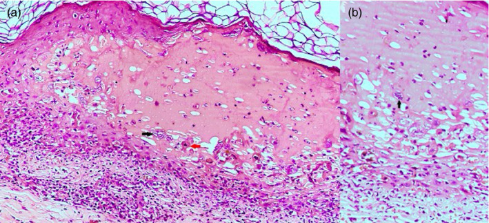
(A): Intraepidermal spongiotic vesicle containing acantholytic cells with large vesicular nuclei (black arrow), neutrophils, and dyskeratotic cells (red arrow). (H&E ×200). (B): Occasional multinucleate cell with ground‐glass chromatin and molded nuclei within the blister (H&E ×400)