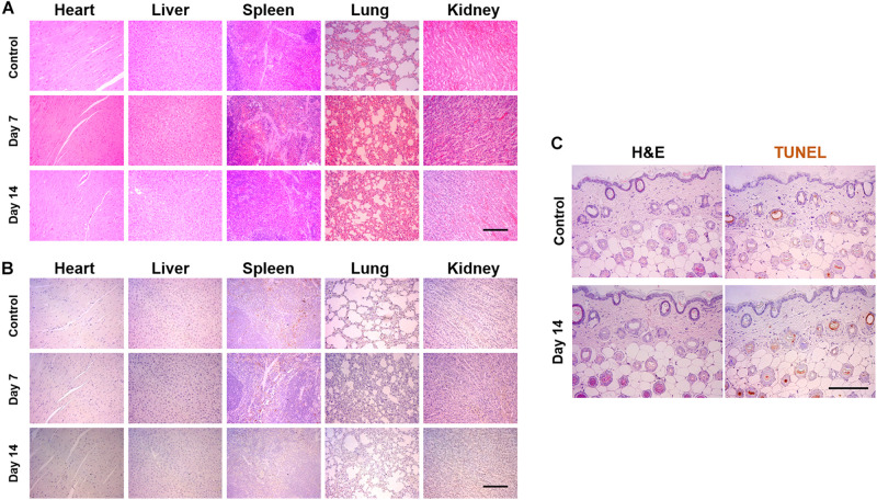
FIGURE 7.
Pathological analysis of SF via intraperitoneal injection and applying to the skin surface. The 50 μl of SF was injected every other day until 2 weeks or applied to the dorsal skin surface of mice. (A) Hematoxyline and eosin (H&E) and (B) TUNEL staining of the tissues harvested from the heart, the liver, the spleen, the lung, and the kidney. (C) H&E- and TUNEL-stained images of the skin, which did not display any toxical responses (Scale bar = 200 μm).

