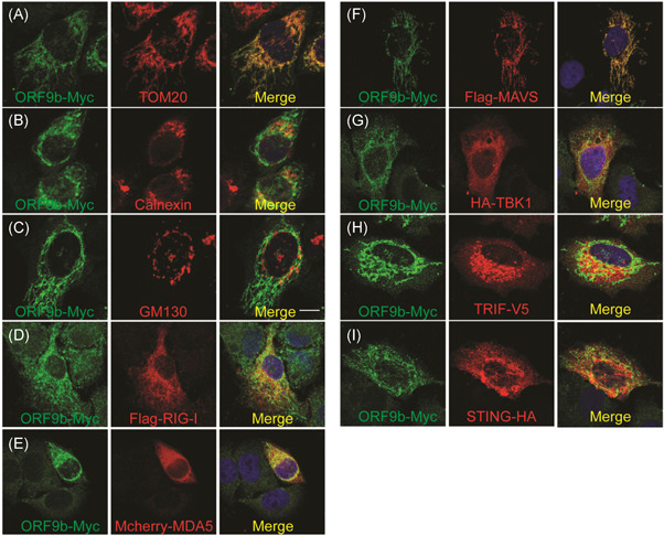
Figure 4.

(A–C) Subcellular localization of SARS‐CoV‐2 ORF9b. HeLa cells seeded on 12‐well coverslips were transfected with the indicated plasmids. Twenty hours after transfection, the cells were subjected to immunofluorescence staining with mouse anti‐Myc antibody and rabbit antibodies against the corresponding organelle marker. Scale bar = 10 μm. (D–I) Relative localization of SARS‐CoV‐2 ORF9b protein with signaling molecules, including RIG‐I, MDA5, MAVS, TBK1, TRIF, and STING. The seeding and transfection of HeLa cells were performed as described in (A). After transfection, ORF9b was stained with a rabbit anti‐Myc antibody, and the signaling molecules were reacted with mouse antibodies against the indicated tags. Scale bar = 10 μm. TOM20, Mitochondria marker; Calnerxin, ER marker; GM130, Golgi marker. SARS‐CoV‐2, severe acute respiratory syndrome coronavirus 2; TBK1, TANK‐binding kinase 1; TRIF, TIR‐domain‐containing adapter‐inducing interferon‐β
