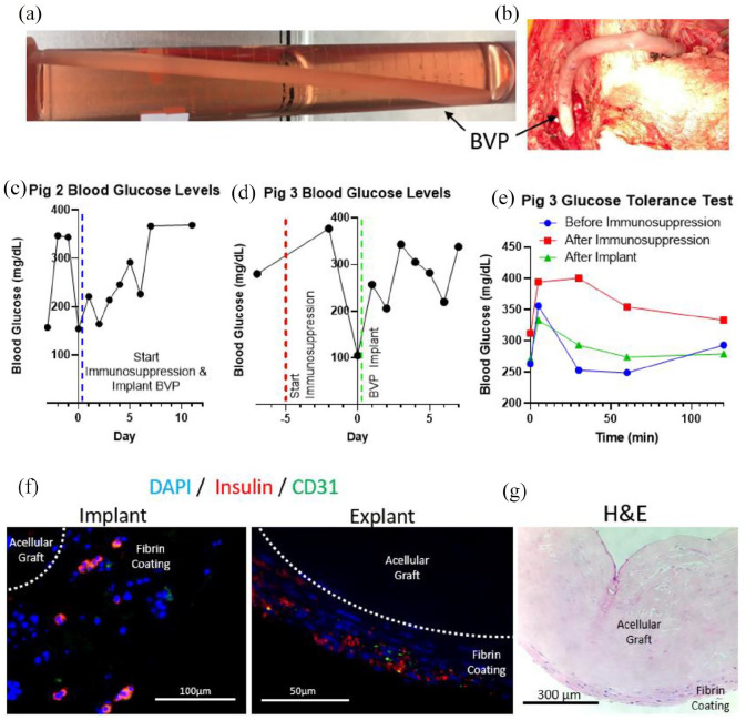
Figure 5.
Scaled-up BVP implantations into immunosuppressed, streptozotocin induced diabetic pigs: (a) BVP 20 cm in length with an inner diameter of 6 mm and outer diameter of 8 mm, (b) BVP implanted as a side-to-side arteriovenous graft from the right common carotid artery to the left external jugular vein, (c) blood glucose levels for second pig that received a BVP implant, (d) blood glucose levels and (e) glucose tolerance test for third pig that received a BVP implant, (f) implant with pancreatic islet fragments containing beta cells in the implant (left) and explant (right), and (g) H&E image of explanted BVP demonstrating no fibrosis on the BVP surface.

