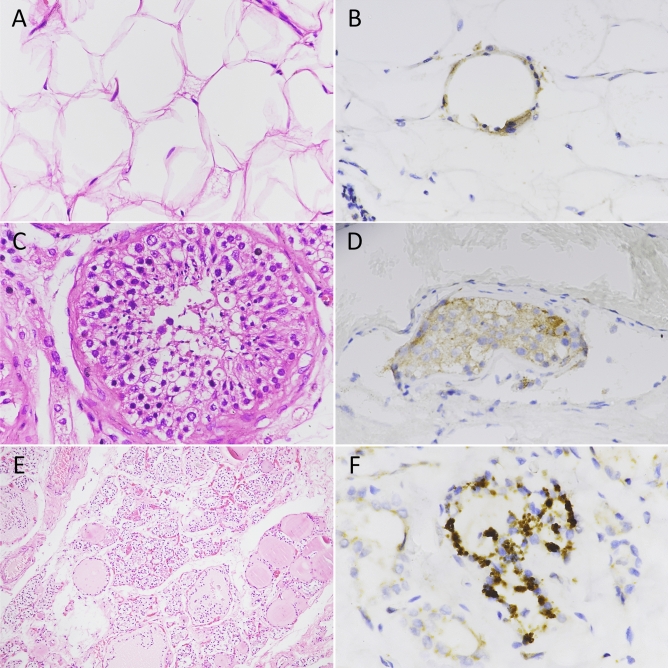
Fig. 2.
Hematoxylin and eosin staining of adipose, testis, and thyroid specimens (A, C, E) and immunohistochemical staining of SARS-CoV-2 nucleocapsid antigen (B, D, F). A Histologic sections of adipose tissue showing adipocytes of different size. B Granular distribution of SARS-CoV-2 nucleocapsid antigen in adipose tissue. C High magnification of normal testis showing seminiferous tubuli. D Moderate brown staining of SARS-CoV-2 nucleocapsid antigen in the cytoplasm of testis cells. E Low magnification of thyroid parenchyma. Follicles of different size are present. F Strong cytoplasmic staining for SARS-CoV-2 nucleocapsid antigen in thyroid follicular cells. Original magnification: A 40 × , B 40 × , C 40 × , D 40 × , E 10 × , F 60 ×

