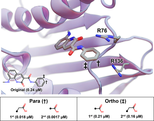
Figure 3.
An inhibitory scaffold bound to E. coli DNA gyrase B. The protein is shown as a blue ribbon, and key amino acids are shown as thin sticks (PDB ID 6KZZ(17)). The scaffold is shown as thick sticks, with the relevant atomic coordinates taken from the 6KZZ ligand.17 The benzene para and meta positions are marked with a dagger and a double dagger, respectively. A 2D depiction of the scaffold is overlaid, with its experimentally measured IC50 value (per Ushiyama et al.17). DeepFrag-suggested fragment additions at the para and ortho positions are ranked in the table below, with the associated IC50 values.

