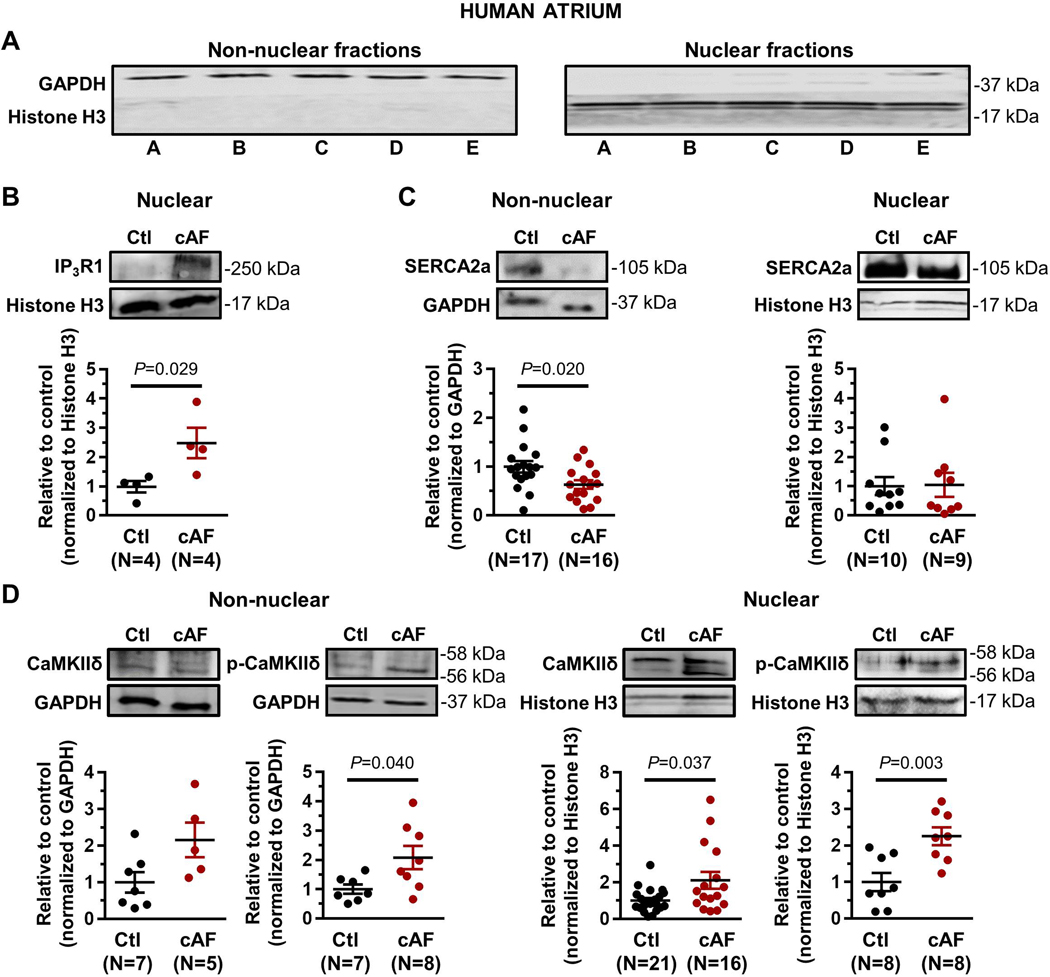
Figure 7.
A. Western blot showing separation of nuclear and non-nuclear fractions from human atrial cardiomyocytes. B. Western blots and quantification of nuclear IP3R1 (Ctl=sinus-rhythm control samples; cAF=chronic AF patient samples). No IP3R1-signal could be detected in the non-nuclear fraction. C. Western blots and quantification of SERCA2a in subcellular fractions. D. Western blots and quantification of total CaMKIIδ and p-CaMKIIδ (Thr287) in subcellular fractions. P-values reflect Ctl vs. cAF, based on Student’s non-paired t-test (non-nuclear SERCA2a in panel C, nuclear p-CaMKIIδ in panel D) or Mann-Whitney test (all other comparisons). Histone H3 and GAPDH were loading controls.

