
Figure 3.
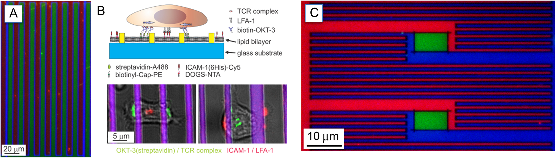
(A) Example of interdigitating lipid bilayers. A serpentine barrier of photoresist (blue) separated bilayers containing a small amount of TR (red) or NBD (green) labeled lipids. (B) Schematic of the use of these segregated bilayers to spatially control the distribution of membrane-tethered anti-CD3 (OKT3, green) and ICAM-1 (red). (C) A finer three-component pattern with submicrometer resolution was achieved using e-beam lithography. A more complicated barrier geometry allowed patterning of an increasing number of proteins. This surface was exposed to lipid vesicles containing small amounts of lipid labeled with TR (red), NBD (green), or DiD (blue). Reprinted with permission from (Shen, Tsai, Shi & Kam, 2009). Copyright 2009 American Chemical Society.