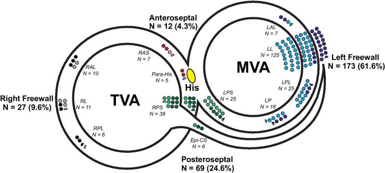
Fig. 2.
Distribution of accessory atrioventricular pathways. The number and proportion of patients with accessory AP locations are displayed along tricuspid valve annulus (TVA) and mitral valve annulus (MVA). His bundle recording is labeled yellow. Number of patients are proportionately represented by the number of circles (each circle represents 2 patients). Anteroseptal pathways are red (manifest) or pink (concealed). Left freewall pathways are dark blue (manifest) or light blue (concealed). Posteroseptal pathways are dark green (manifest) or light green (concealed). Right freewall pathways are black (manifest) or gray (concealed). Sub-regional locations are listed. Epi-CS epicardial-coronary sinus, LAL left anterolateral, LL left lateral, LP left posterior, LPL left posterolateral, LPS left posteroseptal, RAL right anterolateral, RAS right anteroseptal, RL right lateral, RP right posterior, RPL right posterolateral, RPS right posteroseptal

