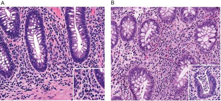
FIGURE 2.
Representative images of basal plasmacytosis (A) and active histological inflammation (B). The inset in A is a higher-power image of plasma cells in the lamina propria adjacent to a crypt base. In B, the main image shows cryptitis (neutrophil infiltration of crypt epithelium), and the inset shows a crypt abscess.

