Fig. 1.
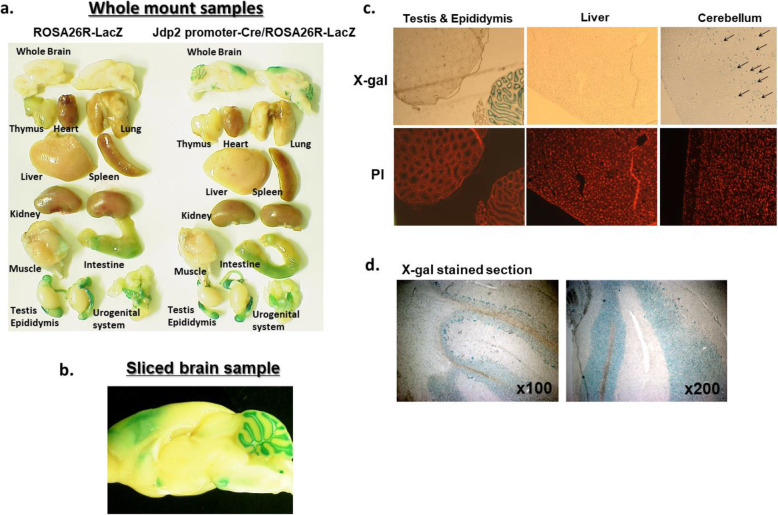
Development of cerebellar lobes and granule cells in Jdp2-promoter-Cre mice. a β-Gal staining of the Jdp2 promoter-Cre/ROS26R-LacZ adult mouse revealed signals in the various organs. WT 129-C57/BL6J ROSA26R-LacZ and Jdp2-promoter-Cre/ROSA26R-LacZ male mice were compared (8–22 weeks), and the brain displayed strong β-Gal staining in WT 129-C57/BL6J Jdp2-promoter-Cre/ROSA26R-LacZ male mice at P24. b β-Gal staining was mainly detected in the cerebellum and some parts of the cerebrum of WT 129-C57/BL6J Jdp2-promoter-Cre/ROSA26R-LacZ male mice at P24. c The β-Gal staining was detected in testis, epididymis, and cerebellum, but not in the liver in WT 129-C57/BL6J Jdp2-promoter-Cre/ROSA26R-LacZ male mice at P24. Arrows indicate β-Gal-stained cells. Propidium iodide (PI) was used as a nuclear DNA counterstain. d β-Gal-stained cells in the cerebellum from WT 129-C57/BL6J Jdp2-promoter-Cre/ROSA26R-LacZ male mice at P24 were viewed under bright field at × 100 and × 200 magnification. The data were shown as one example among 9 male mice at P24

