Fig. 5.
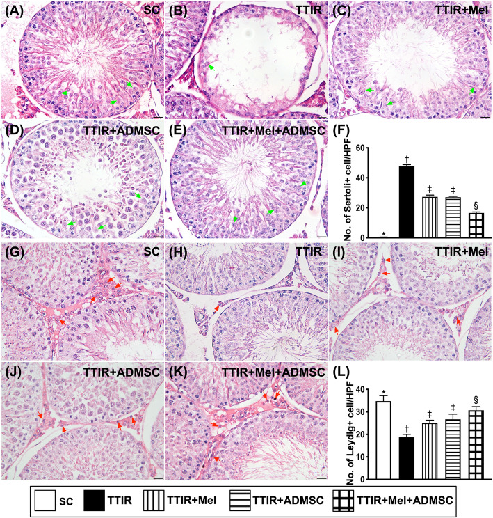
Mel-ADMSCs therapy effectively protected Sertoli cells and Leydig cells against TTIR injury by 72 h after IR procedure. A–E Illustrating the microscopic (× 400) finding of H&E stain for identification of histological feature (i.e., cross-section) of Sertoli cells (green arrows). The result showed that number of Sertoli cells was notably reduced in TTIR groups than in other groups. F The analytical result of the number of Sertoli cells, * vs. other groups with different symbols (†, ‡, §), p < 0.0001. G–K Illustrating the microscopic (× 400) finding of H&E stain for identification of histological feature (i.e., cross-section) of Leydig cells (red arrows). The result demonstrated that number of Leydig cells was notably reduced in TTIR groups than in other groups. L The analytical result of the number of Leydig cells, * vs. other groups with different symbols (†, ‡, §), p < 0.0001.
HPF, high-power field. All statistical analyses were performed by one-way ANOVA, followed by Bonferroni multiple comparison post hoc test (n = 6 for each group). Symbols (*, †, ‡, §) indicate significance (at 0.05 level)

