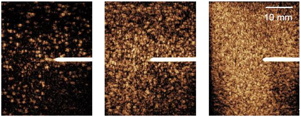
Figure 1:

Contrast-enhanced ultrasound images with the L9-3 linear array of the Philips iU22 showing the dilution of microbubbles in egg white phantoms at three different microbubble concentrations: (a) low (~102 MBs/mL), (b) medium (~103 MBs/mL), and (c) high (~104 MBs/mL). Thermocouples (white lines centered vertically) were inserted into each phantom to measure the temperature rise during each thermal treatment.
