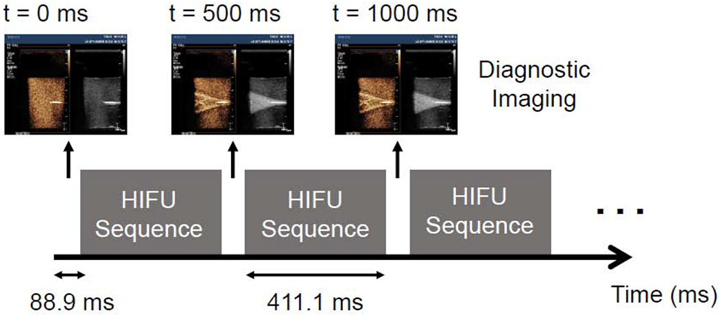
Figure 3:

Schematic showing the timing of the HIFU pulses and diagnostic imaging. Diagnostic imaging was interspersed with the HIFU pulse to allow real-time visualization of the treatment. The therapy transducer is turned on for 370,000 cycles (411.1 milliseconds) followed by a period where an ultrasound image is taken (88.9 milliseconds); this is repeated throughout the entire 30 second treatment.
