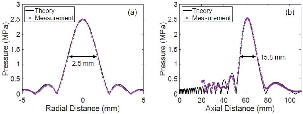
Figure 4:

Verification of the focal spot of the HIFU transducer. (a) Beam pattern taken at the focus of the transducer. The beam width (defined by the −6 dB points) at the focus is 2.5 mm. (b) Axial pressure field of the transducer. The axial beam width is 15.4 mm.
