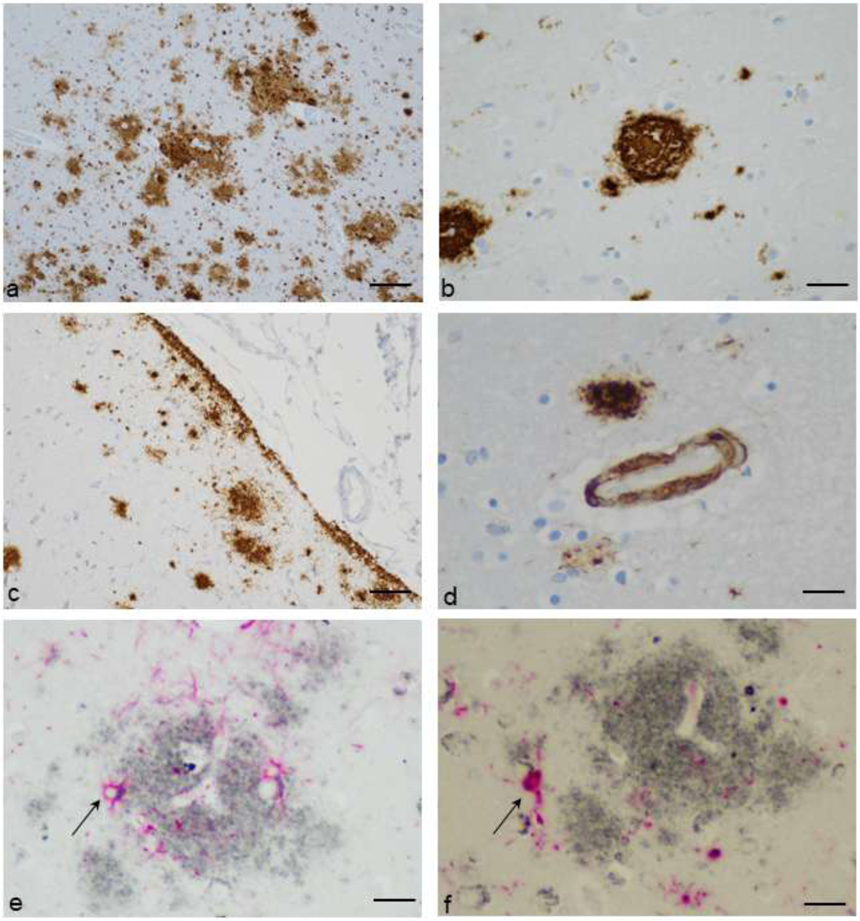
Figure 2.

Patterns of Aβ deposition. a. Diffuse amyloid plaques of varying sizes in cerebral cortex, with and without paravascular locations (Aβ immunostain with diaminobenzidine chromogen and hematoxylin counterstain, original magnification 100X, scale bar = 100um). b. A dense core plaque (Aβ immunostain with diaminobenzidine chromogen and hematoxylin counterstain, original magnification 400X, scale bar = 25um). c. Subpial, diffuse deposits of amyloid (Aβ immunostain with diaminobenzidine chromogen and hematoxylin counterstain, original magnification 200X, scale bar = 50um). d. Cerebral congophilic angiopathy, with adjacent diffuse plaques and glial Aβ (Aβ immunostain with diaminobenzidine chromogen and hematoxylin counterstain, original magnification 600X, scale bar = 16um). e. Aβ plaque (gray) and reactive astrocytes identified with immunohistochemical stain for glial fibrillary acidic protein (GFAP) (red). Aβ is seen within astrocyte cytoplasm (representative arrow). (Immunohistochemical stain for Aβ and GFAP with ImmPACT SG and ImmPACT Vector Red chromogens, original magnification 600X, scale bar = 16um). f. Aβ plaque (gray) and reactive microglia identified with immunohistochemical stain for Iba1 (red). Aβ is seen within microglial cell cytoplasm (representative arrow). (Immunohistochemical stain for Aβ and Iba1 with ImmPACT SG and ImmPACT Vector Red chromogens, original magnification 600X, scale bar = 16um).
