Fig. 7.

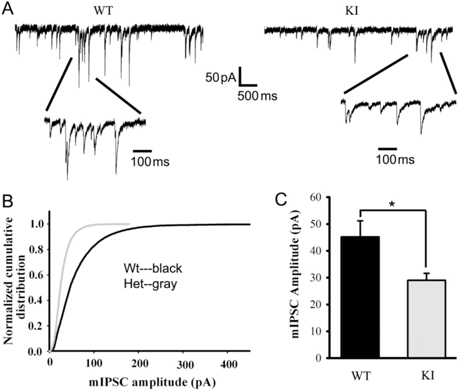
mIPSCs recorded from Gabrg2+/K328M KI mouse SS cortex layer V/VI neuron were altered. A. mIPSCs were recorded from SS cortex layer V/VI neurons (voltage-clamped at −60 mV with equal chloride concentration inside and outside cells) in thalamocortical slices from KI mice and WT littermates. The ACSF perfusion solution contained 20 μM NBQX and 1 μM TTX. B. Normalized cumulative distribution was plotted for WT and KI mouse mIPSCs. C. mIPSCs recorded from SS cortex layer V/VI neurons of WT mice were compared to those in KI mice, showing significantly reduced amplitudes (WT 45.11 ± 6.07 pA, n = 4 mice; KI 28.98 ± 2.62 pA, n = 5 mice. Data were presented as mean ± SEM. Student’s t-test p = 0.036).
