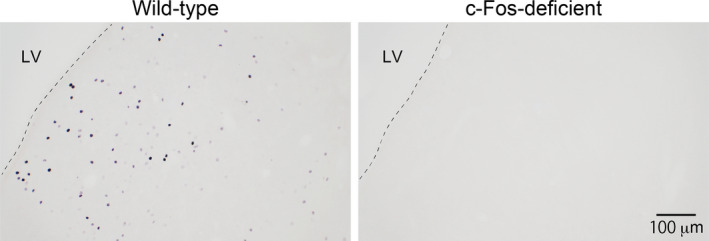
FIGURE 3.

No detection of c‐Fos protein immunoreactivity in brain sections of c‐Fos‐deficient mice. Photographs of sections containing the septum region are shown. Brain sections were processed for detection of c‐Fos protein as described in the Materials and methods. No c‐Fos protein‐immunoreactive profiles (black nuclear profiles) were observed in sections of c‐Fos‐deficient mice. The numbers of wild‐type and c‐Fos‐deficient mice were 3 and 5, respectively. LV, lateral ventricle. Scale bar = 20 μm
