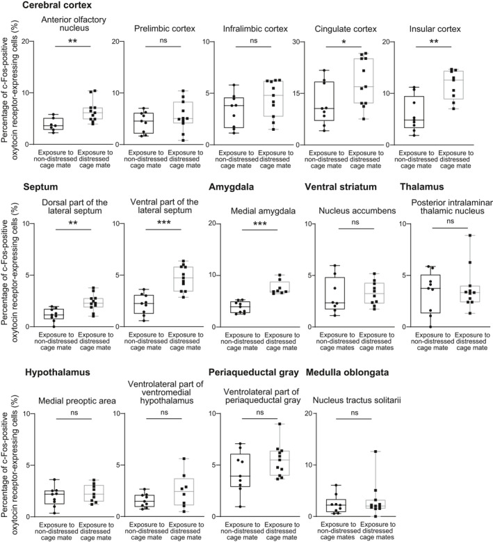
FIGURE 4.

Percentages of oxytocin receptor‐expressing neurones expressing c‐Fos immunoreactivity after exposure to distressed cage mates in oxytocin receptor‐Venus knock‐in heterozygous female mice. Oxytocin receptor‐expressing cells were detected by Venus immunoreactivity in oxytocin receptor‐Venus knock‐in mice. Exposure to distressed cage mates activated oxytocin receptor‐expressing neurones in the anterior olfactory nucleus, cingulate cortex, insular cortex, lateral septum and medial amygdala. The numbers of animals in the groups for non‐distressed control cage mates and for distressed cage mates were 7‐9 and 8‐11, respectively. The maximum, upper quartile, median, lower quartile and minimum values are shown. *P < 0.05, **P < 0.01, ***P < 0.001 vs non‐distressed control cage mates (Mann‐Whitney U test). NS, not significant
