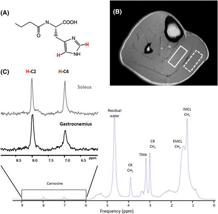
FIGURE 8.

1H spectrum of the carnosine. (A) Chemical structure of carnosine. (B) Positioning the voxel for 1H MRS carnosine acquisition in soleus (full line box) and gastrocnemius (dashed line box) at 3 T using a PRESS sequence and a 15.5 cm diameter, 15‐channel, Tx/Rx extremity coil. Voxel size: 40 mm x 12 mm x 30 mm; TR: 2000 ms; TE: 30ms; 128 averages. Imidazole resonances acquired from the soleus (lower panel, C). The inherent linewidth of the H‐C2‐carnosine peak is ~ 6.5 Hz. Left insert (C) compares the 1H spectrum of the H‐C2 and H‐C4‐carnosine imidazole resonances acquired from the soleus and gastrocnemius at 3 T. The effects of residual dipolar coupling are negligible in soleus due to optimal muscle fiber alignment, resulting in equivalence of the H‐C2 and H‐C4‐carnosine resonances. In gastrocnemius, fiber‐alignment is nonoptimal and the H‐C4‐carnosine peak exhibits dipolar‐coupling induced splitting, which, along with muscle‐specific differences in T2, contribute to the signal mismatch
