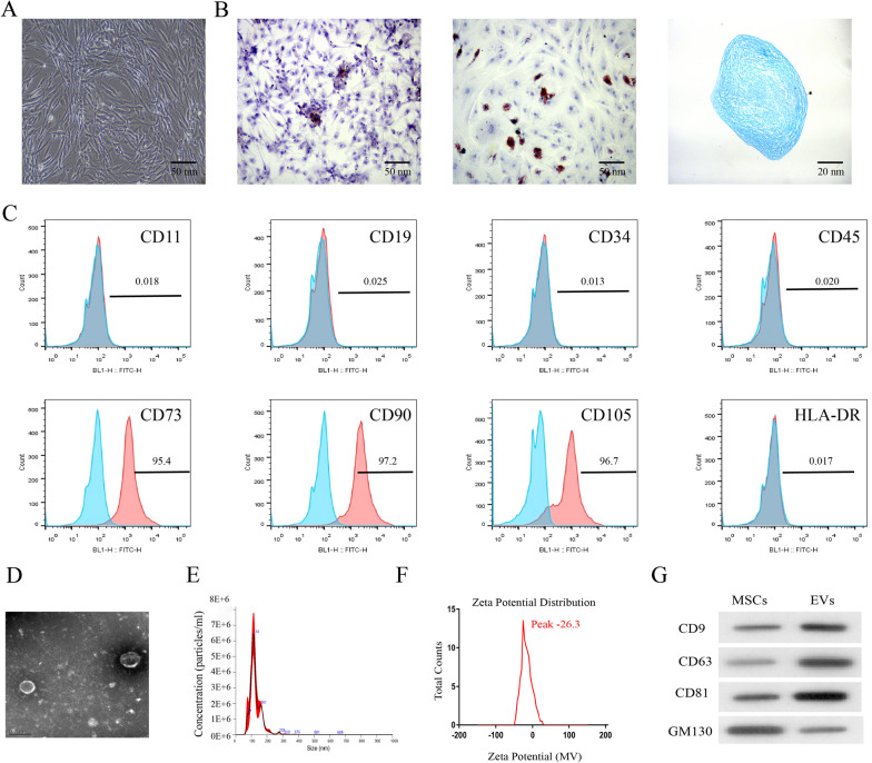
Fig. 1.
Isolation and characterisation of MSC-derived EVs. A After the culture of mesenchymal stem cells (MSCs), microscopic observation showed a typical spindle shape of cells. B Extracted MSCs had osteogenic, adipogenic and chondrogenic potentials by Alizarin Red, Oil Red O and Alcian Blue staining. C MSC positive markers (CD73, CD90 and CD105) and negative markers (HLA-DR, CD11b, CD19, CD34 and CD45) were analyzed by flow cytometry. D The morphology of EVs was observed under a transmission electron microscope. E The particle size distribution of MSC derived EVs was measured by nanometer size analyzer. F Zeta potential measurements of the surface charge of MSCs-EVs (mV). G EVs markers (CD9, CD63, CD81 and GM130) were analyzed by Western blot

