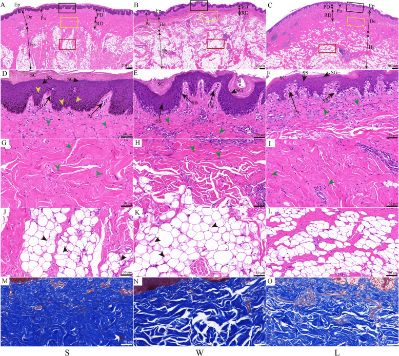
Fig. 1.
Histology of skin located on knob between knob- and non-knob geese. A-C Low-magnification photomicrograph of HE-stained skin. Ep, epidermis; De, dermis; Hy, hypodermis; Pa, papillary; PD, papillary dermis; RD, reticular dermis; the black, yellow and red rectangles represented D-F, G-I, and J-L at high-magnification, respectively; Scale bar: 200 μm. D-L High-magnification photomicrograph of HE-stained skin. SC, stratum corneum; SG, stratum granulosum; SS, stratum spinosum; SB, stratum basal; yellow arrowhead, pigment particles; green arrowhead, fibroblast; black arrowhead, locular adipocytes. Scale bar: 50 μm. M-O High-magnification photomicrograph of Masson-stained skin. Scale bar: 50 μm. S, Lion head goose; W, Sichuan White goose; L, Landes goose. S and W are with knob, L is devoid of knob. The first column represented S, the second column represented W, and the third column represented L

