Fig. 2.
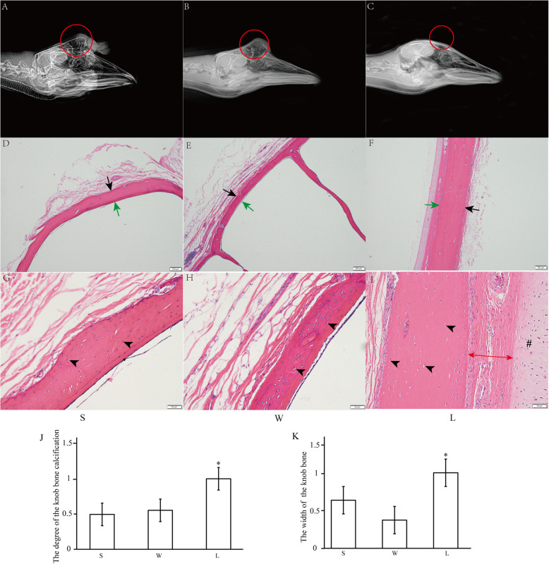
Histomorphology of bone located in knob between knob- and non-knob geese. A-C Right latero-lateral radiograph images of heads of different geese breeds. The position of bone protuberances was red circled. D-F Low-magnification photomicrograph of HE-stained bone. Black arrow, external periosteum; green arrow, internal periosteum. Scale bar: 200 μm. G-I High-magnification photomicrograph of HE-stained bone. Black arrowhead, osteocyte; double-arrow, the connective tissue; #, cartilage tissue. Scale bar: 50 μm. J-K The calcification degree and width of the knob bone in different geese breeds. The data of L was used for normalization of the data of both S and W, and the data was displayed as multiples of changes. * represented the statistically significance. P < 0.05. S, Lion head goose; W, Sichuan White goose; L, Landes goose. S and W are with knob, L is devoid of knob

