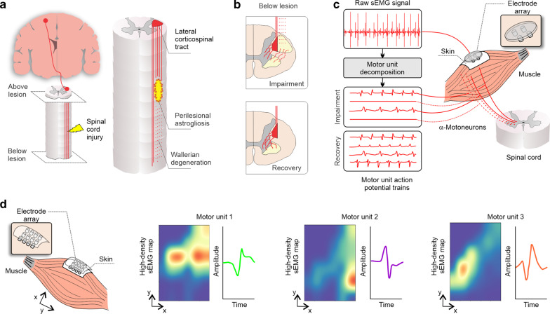
Fig. 8.
Overview of conceptual work on high-density sEMG, motor unit decomposition, and its application in SCI. a, left Corticospinal projections (red) and spinal cord injury (yellow). a, right In the intermediate and chronic phases (2 weeks to 6 months), axons continue to degenerate and the astroglial scar matures to become a potent inhibitor of regeneration (restrict axonal regrowth and cell migration). The lateral corticospinal tract (red) is the major descending motor tract, which may be damaged after SCI (red hashed lines) [192]. b, upper panel The remaining projections from the corticospinal tract (red solid lines) synapse with α-motoneurons in the spinal cord to control volitional movements—(b, lower panel) which undergo extensive plasticity with motor recovery [193]. c Standard sEMG is able to capture the overall activity of these motor units but four or five small pin electrode arrays are able to decompose the raw sEMG signal into individual motor unit potential trains [186, 194, 195], with potential to track the impairment and recovery of motor unit control after an SCI. The most common techniques used for motor unit decomposition involve the use of the progressive FastICA peel-off framework [196–198], multichannel blind source separation using convolution kernel compensation [199–201] or specific algorithms, e.g., using machine-learning and time-varying shape discrimination [186, 194, 195]. d The use of multi-electrode arrays increases the spatial resolution; in addition to the motor unit decomposition, multi-electrode arrays can also unveil the territory of each motor unit [202]

