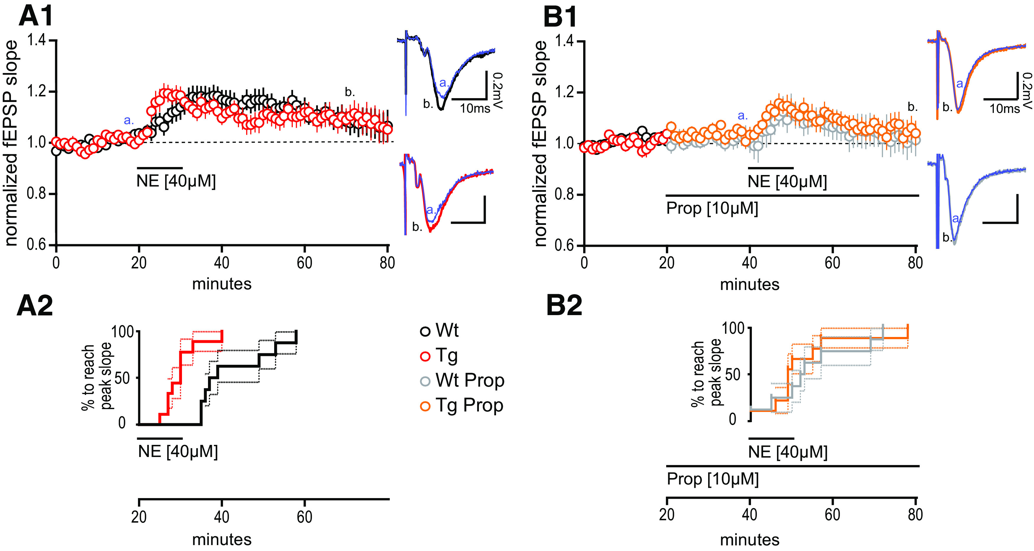
Figure 2.

Heightened synaptic potentiation at MPP–DGC synapses in TgF344-AD rats induced by norepinephrine requires β-adrenergic receptors. A1, NE (40 μm) was bath applied during extracellular dendritic field potential recordings at MPP–DGC synapses in slices from 9- to 10-month-old Tg (n = 10) and WT (n = 8) rats. Representative averaged fEPSP traces (five sweeps) from WT (black) and Tg (red). Calibration: 0.2 mV, 10 mS at times t [a = 20 min (blue); b = 60 min]. Long-term potentiation was reached for Tg fEPSPs at t(65–70), 40 min following NE washout (p < 0.05), and was not different between genotypes (p > 0.1). A2, Survival analysis plot displaying the time at which the fEPSP in each slice reaches peak potentiation, demonstrating a significantly more rapid time to peak in Tg compared with WT (p < 0.001). B1, B2, The β-AR antagonist PROP (10 μm) prevents the rapid increase in synaptic potentiation in Tg rats (p > 0.1; B2), and blocks the long-lasting potentiation in both genotypes from t(35–40) to t(75–80) of Tg (n = 9) and WT (n = 8) when compared with paired t tests (both, p > 0.1). Representative traces for WT + PROP (gray) and Tg + PROP (orange) with calibration (0.2 mV, 10 mS) at times t (a = 40 min; b = 80 min). All values are the mean ± SEM.
