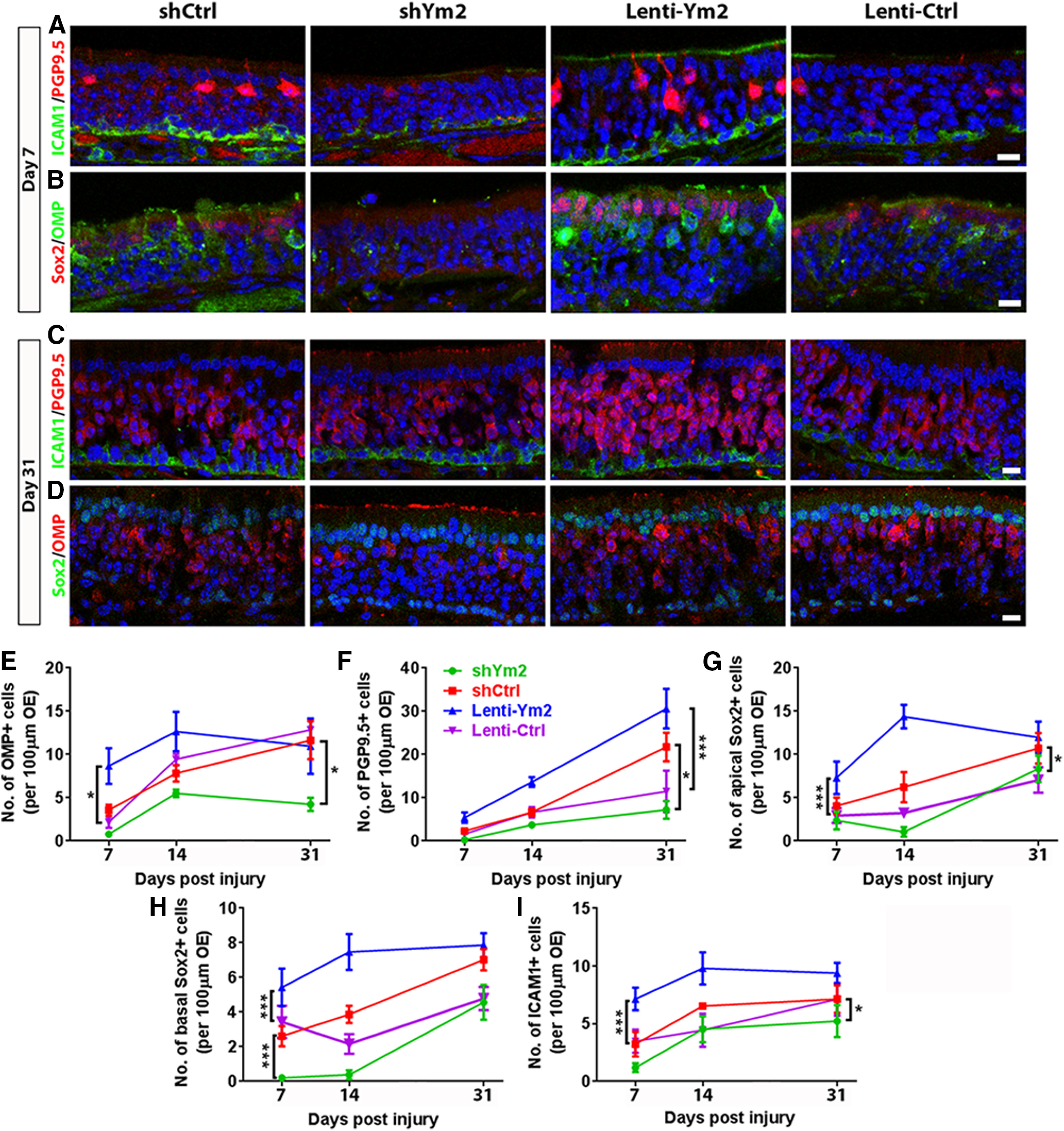
Figure 6.

Ym2 regulates regeneration in the lesioned OE. A–D, Confocal images of ICAM1+, PGP9.5+, OMP+, and Sox2+ cells in the lesioned OE from mice injected with Lenti-shCtrl, Lenti-shYm2, Lenti-Ym2, or Lenti-Ctrl on day 7 and day 31 postinjury. E–I, Statistical analysis on the number of OMP+, PGP9.5+, apical Sox2+, basal Sox2+, and ICAM1+ cells/100 µm OE of mice injected with Lenti-shYm2, Lenti-shCtrl, Lenti-Ym2, or Lenti-Ctrl on day 7, day 14, and day 31 postinjury (OMP: n = 6 sections from three mice in each group; PGP9.5: n = 5–7 sections; apical Sox2+: n = 5–7 sections; basal Sox2+: n = 5–9 sections; ICAM1+: 5–7 sections). Statistical significance was determined by unpaired Student's t test between Lenti-shCtrl and Lenti-shYm2 or between Lenti-Ym2 and Lenti-Ctrl at the same time points, or by two-way ANOVA at all three time points together. Statistical analysis shown in E–I was based on two-way ANOVA. E: F(3,60) = 12.10, p < 0.0001; F: F(3,56) = 16.49, p < 0.0001; G: F(3,59) = 17.9, p < 0.0001; H: F(3,65) = 22.36, p < 0.0001; I: F(3,57) = 12.21, p < 0.0001. Asterisks was determined by Tukey's multiple-comparisons test. Scale bars, 10 μm.
