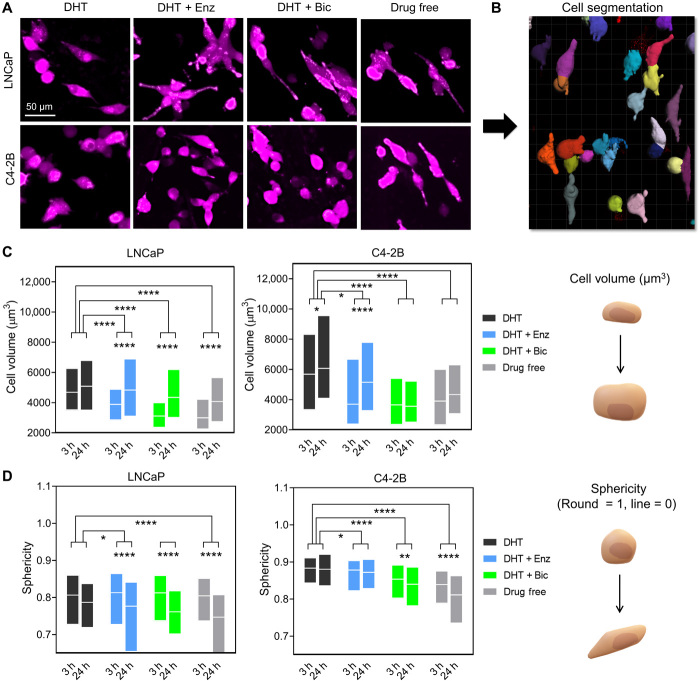
Fig. 2. Antiandrogen treatments increase cancer cell volume and reduces sphericity in the bone microenvironment.
(A) Confocal microscopy images of the 3D metastatic microtissues after 24 hours hOBMT coculture with LNCaP and C4-2B under treatments, showing cancer cell morphology (mKO2) on hOBMT (MaxProj. shown, 70-μm z-stacks). (B) Cell segmentation processing through Imaris software. (C and D) 3D morphometric properties of cancer cells after 3 hours and after 24 hours coculture under treatments, shown as box plots, (C) cell volume, and (D) sphericity (>2 microtissues per condition analyzed with >4 random fields of view, average n = 365 cells). DHT (dihydrotestosterone), 10 nM; Enz (enzalutamide), 10 μM; Bic (bicalutamide), 10 μM. *P < 0.05, **P < 0.01, and ****P < 0.0001.

