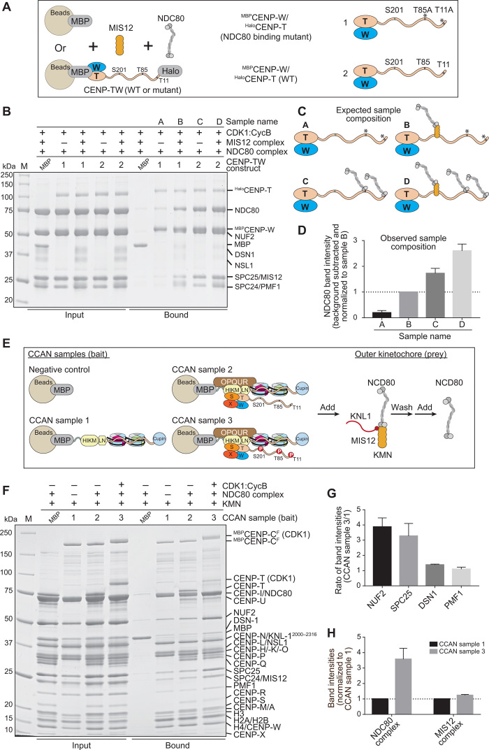
Fig. 8. Reconstitution of a complete human kinetochore module.
(A) Schematic of pull-down experiments. MBPCENP-W/HaloCENP-TWT (1) and MBPCENP-W/HaloCENP-TT11A/T85A (2) immobilized on amylose beads as baits were incubated with NDC80C pray, alone, or with MIS12C. (B) SDS-PAGE result of pull-down experiment. MBPCENP-W/HaloCENP-T bait and added prey are indicated above each lane.( C) Expected composition of samples in (B). (D) Quantified NDC80 band intensities (normalized to sample B) and SDs (three repeats) of samples A to D indicated in bound fractions of (B). NDC80 band intensity of MBP control was subtracted as background. (E) Schematic of pull-down experiment. MBP and three CCAN samples assembled on MBPCENP-CF were immobilized on amylose resin as bait. KMN complex was added and incubated for 1 hour. Unbound KMN was washed, and NDC80C added for 1 hour. After washing unbound NDC80C, samples were analyzed by SDS-PAGE. (F) SDS-PAGE result of pull-down experiment. The CCAN bait and presence of CDK1:Cyclin B are indicated above each lane. (G) Quantified CCAN sample 3/sample 1 ratio and SD (three repeats) of indicated proteins shown in bound fractions of (F). (H) Quantified intensities and SD (three repeats) of the NDC80C (averaged over the NUF2 and SPC25 band intensities) and the MIS12C (averaged over the DSN1 and PMF1 band intensities) normalized to CCAN sample 1.

