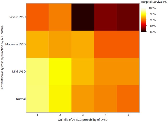
Figure 3.
Heat map demonstrating hospital survival (A) and 1-year survival (B) as a function of LVSD on TTE based on current ASE guidelines16 (Y axis) and AI-ECG probability of LVSD quintile (X axis). Darker colours represent a higher risk of hospital death. AI-ECG, artificial intelligence-augmented electrocardiogram; LVSD, left ventricular systolic dysfunction; TTE, transthoracic echocardiogram.

