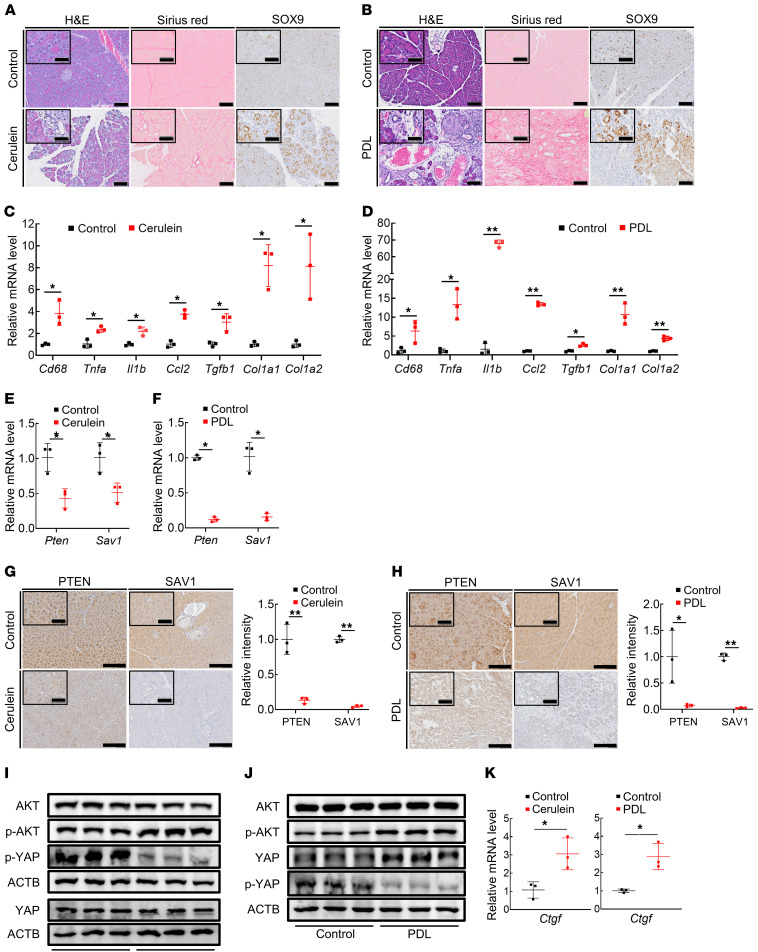
Figure 1. Expression of PTEN and SAV1 is downregulated in the pancreatic tissues of mice in 2 models of CP.
(A and B) Representative images of H&E, Sirius red, and SOX9 staining of pancreatic tissue in mice after repeated cerulein or vehicle (control) injection (A) and in mice subjected to pancreatic duct ligation (PDL) surgery or sham surgery (B). (C and D) Cd68, Tnfa, Il1b, Ccl2, Tgfb1, Col1a1, and Col1a2 mRNA levels in pancreatic tissue in mice after repeated cerulein injection (C) and in mice subjected to PDL surgery (D). (E and F) Pten and Sav1 mRNA levels in pancreatic tissue in mice after repeated cerulein injection (E) and in mice subjected to PDL surgery (F). (G and H) Representative images of PTEN and SAV1 staining of pancreatic tissue in mice after repeated cerulein injection (G, left), with quantification of the PTEN and SAV1 staining intensity (G, right); and in mice subjected to PDL surgery (H, left), with quantification of PTEN and SAV1 staining intensity (H, right). (I and J) Protein levels of AKT, p-AKT, YAP, p-YAP, and ACTB in the pancreata of mice after repeated cerulein injection (I) and in mice subjected to PDL surgery (J). (K) Ctgf mRNA levels in pancreatic tissue in mice after repeated cerulein injection (left) and in mice subjected to PDL surgery (right). Blots run in parallel contemporaneously or run at different times with loading control for each gel are shown. All data are presented as the means ± SDs of results for 3 mice per group. Student’s t test was used to evaluate differences between 2 groups. *P < 0.05 and **P < 0.005. Scale bars: 100 μm and 50 μm (insets).

