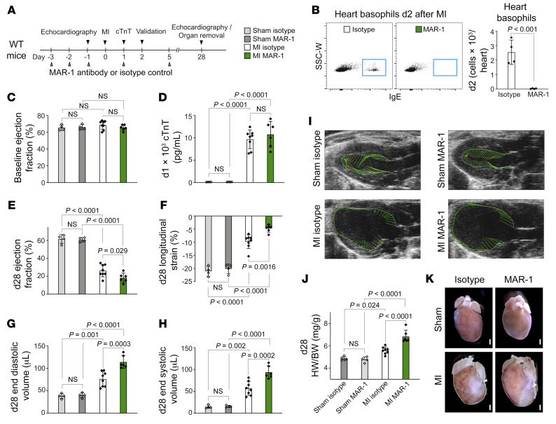
Figure 2. Basophil depletion by antibody injection worsens cardiac function after acute MI in mice.
(A) Timeline of basophil depletion experiments. (B) Frequencies of basophils from of hearts of IgG-injected control and anti-FcεRI–injected animals were assessed by flow cytometry 2 days (d2) after MI (n = 4). Data show the mean ± SD. P value was determined by 2-tailed Student’s t test. (C) Echocardiographic evaluation of baseline EF in IgG- and MAR-1–treated mice. (D) Plasma levels of cTnT in IgG- and MAR-1–treated mice were measured 24 hours after LAD ligation or sham intervention. P values were determined by 2-way ANOVA followed by Tukey’s multiple-comparison test. (E–H) Echocardiographic results for IgG-treated and MAR-1–treated mice 4 weeks after MI or sham surgery. P values were determined by 2-way ANOVA followed by Tukey’s multiple-comparison test. (I) Representative echocardiographic images 4 weeks after MI. Vectors display the direction and magnitude of myocardial contraction at midsystole. (J) Quantification of heart weight to body weight ratio (HW/BW) determined 4 weeks after MI (n = 4–8). Data show the mean ± SD. P values were determined by 2-way ANOVA followed by Tukey’s multiple-comparison test. (K) Representative hearts from IgG- and MAR-1–treated mice 4 weeks after MI or sham intervention. Arrowheads indicate the site of ligation. Scale bars: 500 μm.

