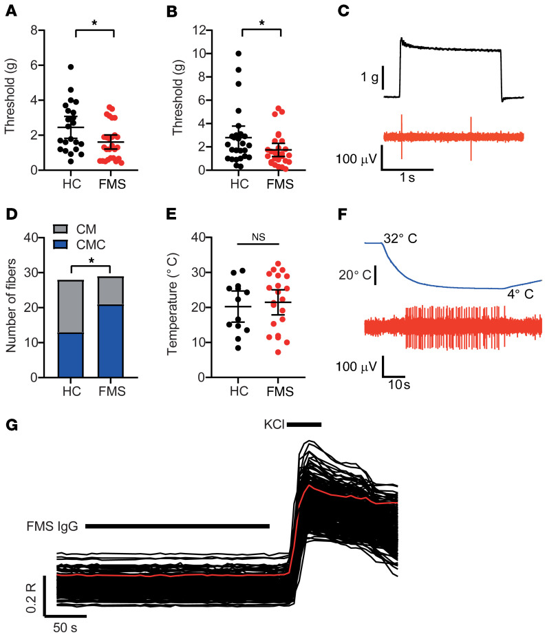
Figure 6. Passive transfer of FMS IgG sensitizes nociceptors.
The mechanical activation thresholds of (A) Aδ- (AM) and (B) C-mechanonociceptors (CM) were reduced in preparations from mice treated with FMS compared with HC IgG (n = 22–27 single units). *P < 0.05 by 1-tailed Mann-Whitney U test. (C) The example trace illustrates a mechanical threshold response (evoked by the minimum force required to elicit at least 2 spikes) in a CM unit. (D) The proportion of cold-sensitive CM units (CMCs) was increased in preparations from mice treated with FMS IgG (21 of 29 units responded to cold) compared with HC IgG (13 of 28 units responded to cold). *P < 0.05 by 1-sided Fisher’s exact test. (E) The cold-activation thresholds of CMC fibers did not differ between FMS and HC preparations (n = 13–21). P > 0.05 by 1-tailed t test. (F) The example trace illustrates a cold-evoked response in a CMC fiber. (G) Application of FMS IgG (200 μg/mL) to isolated DRG neurons loaded with Fura-2 was without effect on [Ca2+]i in all 870 examined neurons (identified by their response to 50 mM KCl). The red trace illustrates the average time course of the displayed 230 neurons.

