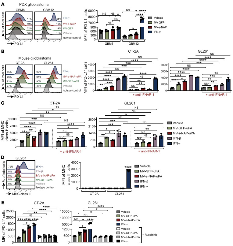
Figure 4. Infection with uPAR-retargeted MV upregulates PD-L1 and MHC class I expression in glioma cells.
CT-2A, GL261, GBM6, and GBM12 cells were infected with the indicated MV strains at MOI = 2. (A) Representative histograms (left) and MFI (right) of PD-L1 surface expression assessed 48 hours after infection in GBM6 and GBM12 PDXs. (B) Representative histograms (left) and MFI (right) of PD-L1 cell surface expression assessed 48 hours after infection in CT-2A and GL261 murine glioma cells. (C) MFI of MHC class I cell surface expression in CT-2A and GL261 glioma cells infected with MV strains in the presence or absence of 10 μg/mL anti–IFNAR-1 depleting antibody. (D) MFI of MHC class II cell surface expression assessed 48 hours after infection in CT-2A and GL261 cells. (E) MFI of PD-L1 surface expression in CT-2A and GL261 glioma cells infected with MV strains in the presence or absence of 5 μM ruxolitinib. Glioma cells were stimulated overnight with 500 IU/mL exogenous IFN-β or IFN-γ to serve as controls. Values represented as the mean ± SD and are representative of at least 2 independent experiments (n = 3 sets of cells/group). *P < 0.05; **P < 0.01; ***P < 0.001; ****P < 0.0001 by 2-way ANOVA with Tukey’s multiple comparison test. NS, not significant.

