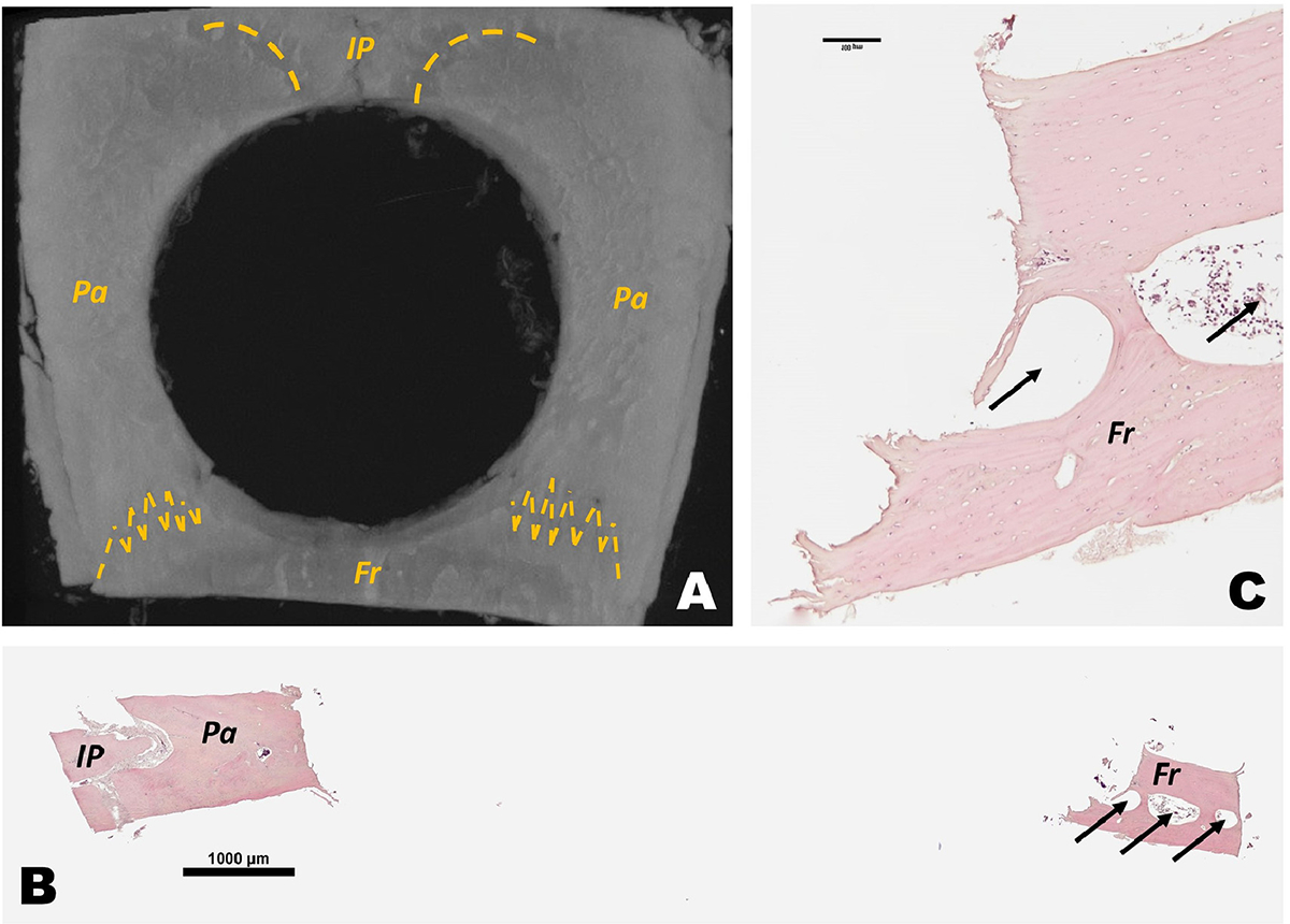
Fig. 4:

Circular critical-sized defect invades frontal and interparietal bone. A) Representative microCT 3D reconstruction of rat calvaria with 7.9mm diameter segmental defect in parietal bones also impacts interparietal and frontal bone at day 0. Yellow dash lines marks the suture between bones. B) H&E histological scan of sagittal sections of circular segmental defect. C) magnified H&E histological sagittal section of frontal bone in a circular segmental defect in calvaria. IP = interparietal bones; Pa = parietal bone; Fr = frontal bones. Arrows indicate frontal bone sinus.
