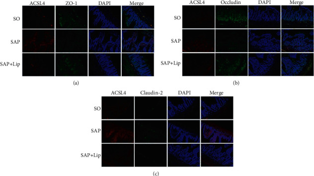
Figure 6.

The expression of ACSL4 and TJ proteins in ileum was observed by confocal laser scanning microscopy. The intestinal sections of SO, SAP, and SAP + Lip groups were double stained with rabbit anti-ACSL4 (red), rabbit anti-ZO-1 (green), rabbit anti-occludin (green), and rabbit anti-claudin-2 (green). The nuclei of the cells were stained with DAPI (blue). (a) ACSL4 (red) and ZO-1 (green), (b) ACSL4 (red) and occludin (green), and (c) ACSL4 (red) and claudin-2 (green) (n = 10, each). Original magnification, ×200.
