Figure 1.

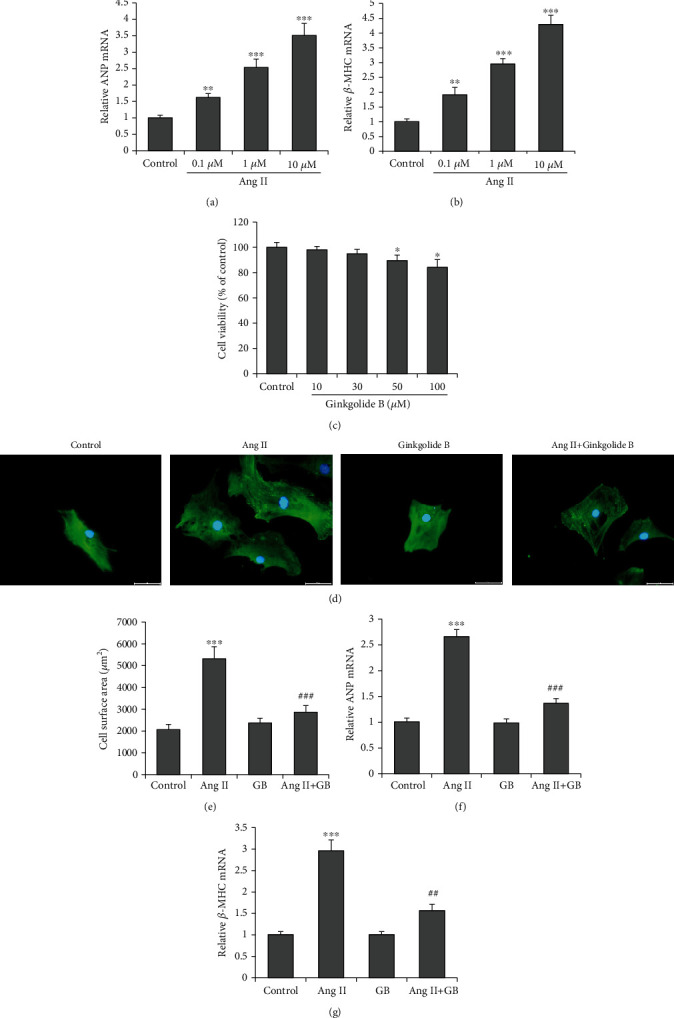
Ginkgolide B attenuates Ang II-induced hypertrophy. H9c2 cells were incubated with Ang II (0, 0.1, 1, and 10 μM) for 48 h. RT-qPCR examined (a) ANP and (b) β-MHC mRNA levels. (c) Cell viability assay (cells were incubated for 48 h) showed the Ginkgolide B with above 50 μM has an obvious toxic effect on cardiomyocytes. (d) H9c2 cells were pretreated with Ginkgolide B (30 μM) for 2 h and then incubated with Ang II (1 μM) for a further 48 h. Representative photographs are shown (scale bar: 5 μm). (e) Cardiomyocyte hypertrophy is evaluated by quantifying cell surface area. mRNA expressions of ANP (f) and β-MHC (g) were evaluated by real-time PCR. Data were presented as the mean ± SD. ∗P < 0.05, ∗∗∗P < 0.001 vs. the control group; #P < 0.05, ##P < 0.01, and ###P < 0.001 vs. the Ang II group.
