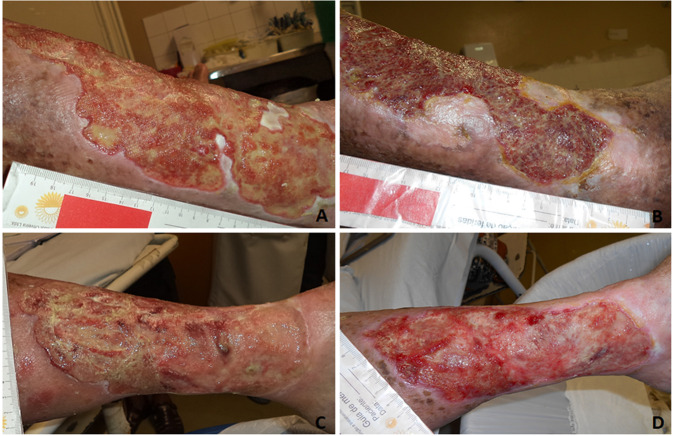
Fig. 3.
Aspect of the chronic venous ulcer. A, B represents the experimental group: A Day 0. Irregular and profound edges, maceration, wound bed with shattered (yemiow) and granulated (red) tissue, large amount of exudate. B After 180 days. Epithelial edges without maceration, presence of granulation and reduction of shattering, increased epithelization areas, low amounts of exudate and macerated islands of epithelial tissue (lateral and inferior edges). C, D represent the control group: C day 0, Irregular edges, maceration, large amount of shattered wound bed (yemiow), large amount of exudate. D Day 180. Epithelialized edge, low amount of maceration on the inferior part, large amount of granulation tissue (red), moderate amount of exudate

