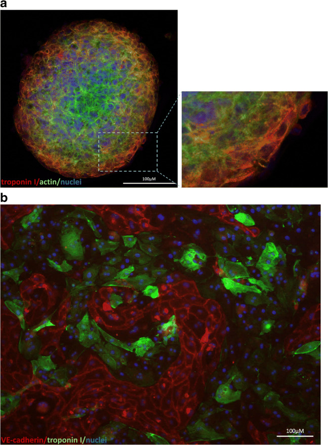
Fig. 3.
hiPSC-derived cardiomyocytes growing in a a spheroid (cells were immunofluorescently stained for troponin I (red) and actin (green); nuclei were stained with 4′6-diamidino-2-phenylindole (DAPI)) and in b co-culture with hiPSC-derived endothelial cells within ibidi μ-slide after 24 h of shear stress (cells were immunofluorescently stained for troponin I (green) (a marker of cardiomyocytes) and VE-cadherin (red) (a marker of endothelial cells); nuclei were stained with DAPI)

