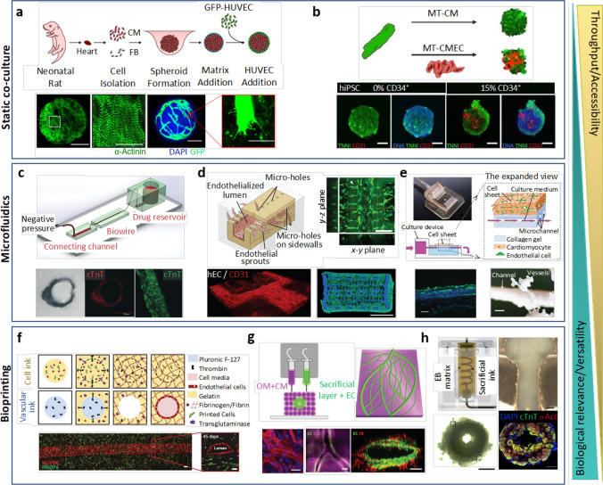
Fig. 2.
In vitro models of myocardial-microvascular interaction. (a) Spheroid based static co-culture of cardiomyocytes (CM), fibroblast (FB), and human umbilical vein endothelial cells (HUVEC) [194]. (b) Co-differentiated human-induced pluripotent stem cell–derived cardiomyocytes (hiPSC-CM)-endothelial cell (EC) 3D microtissue (MT) [47]. (c) Microfluidic Biowire setup used for creation of perfused cardiomyocyte bundle [202]. (d) Microfluidic Angiochip designed to create branching vascular network and endothelialised multi-layer cardiac tissue [212]. (e) Cell sheet technology combined with a perfusion bioreactor creating a vascularized thick tissue [166]. (f) Multi-material extrusion-based bioprinting technique creating thick tissue with vascular lumen [88]. (g) Bioprinted patient-specific thick, perfused, and vascularized cardiac patch made from omental tissue (OM) [136]. (h) Embryoid body (EB)– based bioprinted cardiac tissue using SWIFT method [175]

