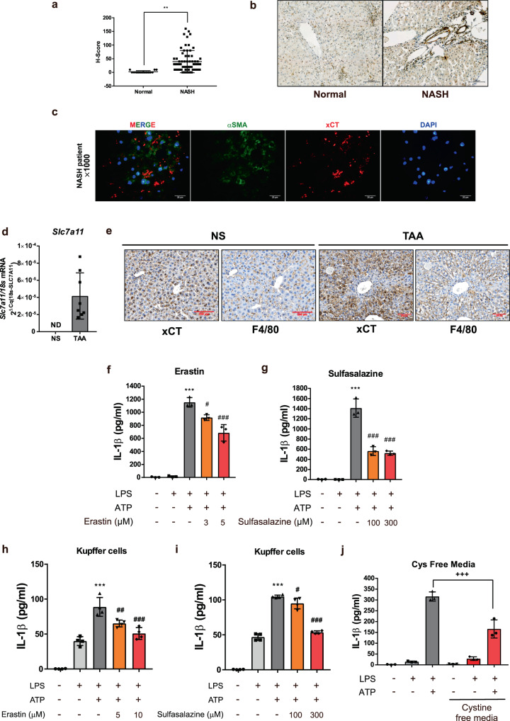
Fig. 8. System Xc is upregulated in profibrotic conditions and its blockade prevents activation of NLRP3 inflammasome.
a H-score based on xCT stained IHC of liver samples from 14 normal and 64 NASH patients. Data are presented as mean ± SD, analyzed by unpaired student t-test; **p < 0.01. b Representative images of xCT stained IHC in normal and NASH liver. Scale bar = 100 µm. c Immunofluorescence images showing the localization of xCT in frozen liver sample from profibrotic NASH patient. Liver tissues were labeled with αSMA (green), xCT (red) and DAPI (blue). Scale bar = 20 µm. d Slc7a11 (xCT) mRNA expression level was measured using real-time qPCR in TAA-induced fibrotic liver (n = 8 mice per group). NS; normal saline, ND; not detected, Slc7a11 Cq value >35. e IHC images showing the localization of xCT in liver samples. Liver tissues were labeled with F4/80 and xCT. NS; normal saline, Scale bar = 200 µm. f, g, and j Effects of erastin, sulfasalazine and cystine free media on ATP triggered IL-1β release of LPS-primed BMDM. h, i Effects of erastin and sulfasalazine on ATP triggered IL-1β release of LPS-primed kupffer cells. Data are presented as mean ± SD (f, g, h, i, and j), analyzed by one-way ANOVA followed by Tuckey’s test. n = 3 for f, g, j and n = 4 for h, i. ***p < 0.001, compared to control; #p < 0.05, ###p < 0.001 compared to LPS, and ATP-treated group; +++p < 0.001, compared to LPS, and ATP-treated group in DMEM media.

