Fig. 1.
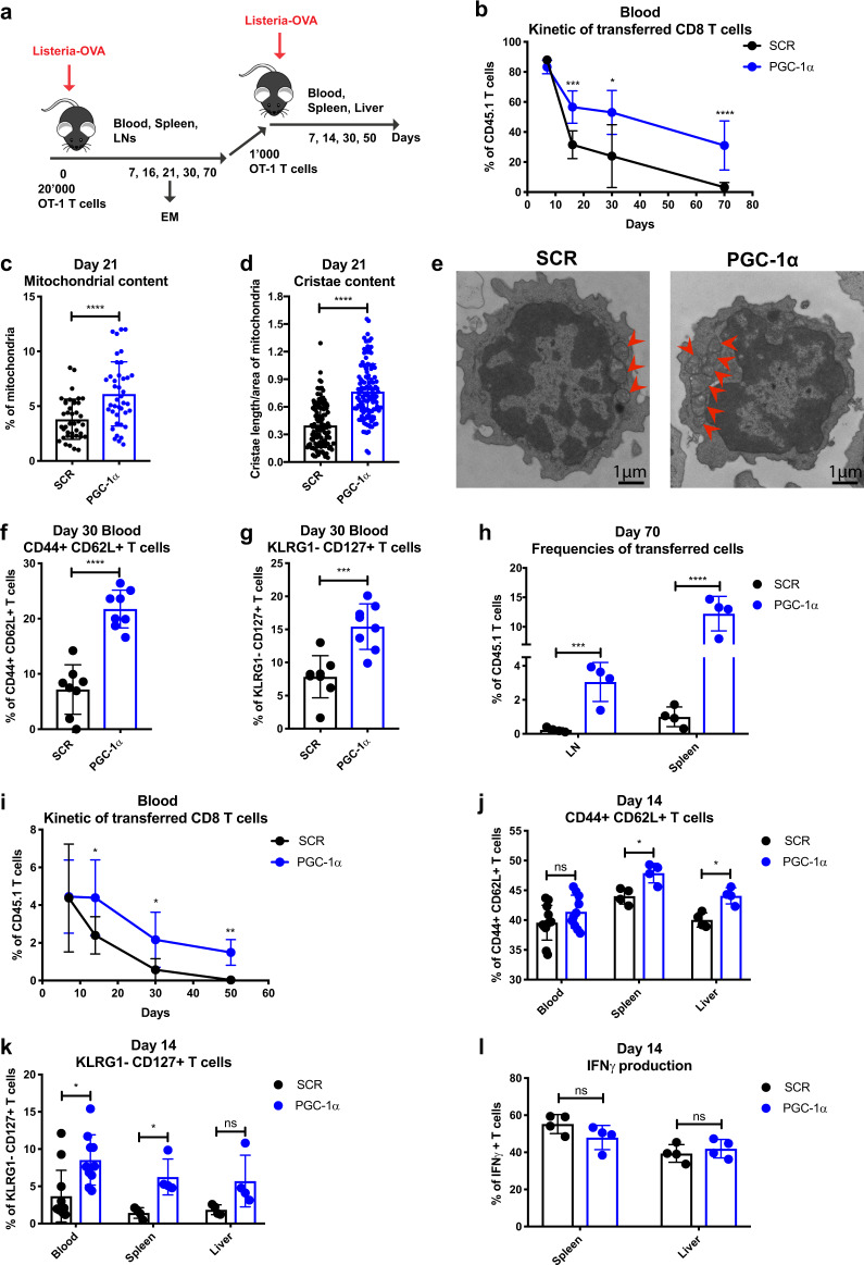
Enforced PGC-1α expression promotes CD8 T cell persistence, memory phenotype, and antigen recall potential. a Schematic representation of Listeria-Ova infection. A total of 20,000 CD45.1 OT-1 cells transduced with PGC-1α or SCR were transferred into CD45.2 naive recipients followed by 2000 CFU of Listeria-Ova. A total of 1000 transduced OT-1 cells were sorted 70 days post infection and transferred to CD45.2 naive hosts rechallenged with 2000 CFU of Listeria-Ova. b Kinetics of the adoptively transferred OT-1 cells in the blood post primary infection. c Percentage of mitochondria per cell area of transduced T cells at day 21 post infection. d Cristae length per area of mitochondria at day 21 post infection. e Representative examples of mitochondrial content per transduced cell. f Percentage in the blood of the CD44+ CD62L+ and g KLRG1− CD127+ populations at day 30 post primary infection. h Frequencies of transferred cells in LNs and spleen on day 70 post primary infection. i Kinetics of the adoptively transferred transduced OT-1 cells in the blood post rechallenge. j Percentage of CD44+ CD62L+ and k KLRG1− CD127+ populations in the blood, spleen, and liver on day 14 post rechallenge. l IFNγ production of transferred T cells in the spleen and liver on day 14 post rechallenge. b, h, i Gated on CD8+, f, g, j–l gated on CD8+ CD45.1+ GFP+. The results are representative of four independent experiments of primary infection and three independent experiments of rechallenge and represent the mean ± SD (4–10 mice per group). c n = 40 cells per group, and d n = 120 mitochondria per group. *p < 0.05; **p < 0.01; ***p < 0.001; ****p < 0.0001

