Fig. 3.
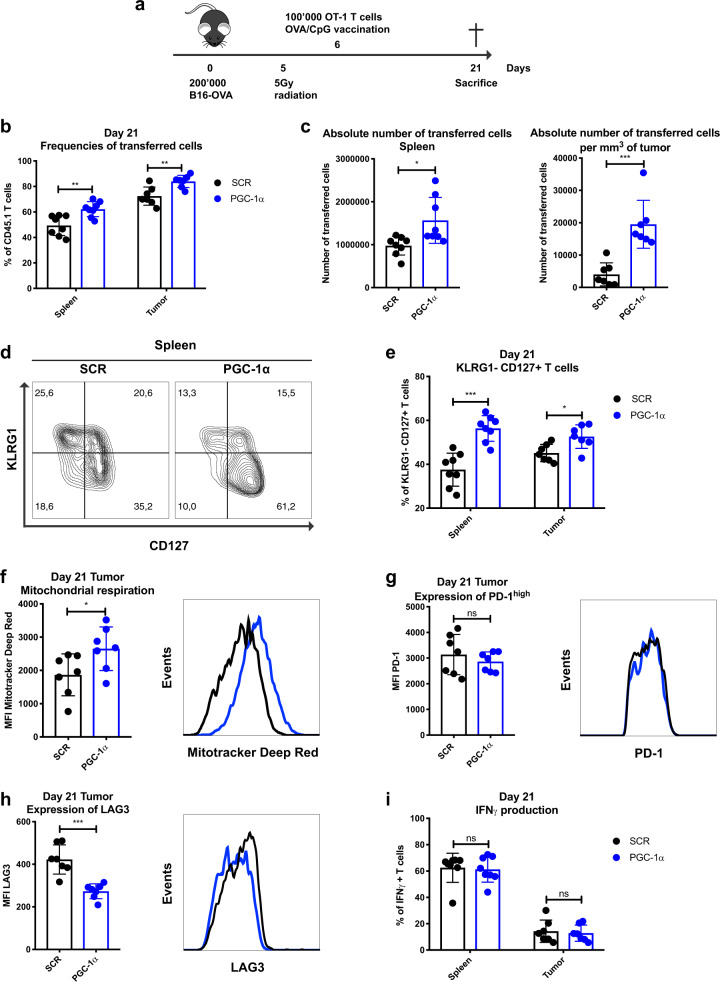
CD8 T cells overexpressing PGC-1α show enhanced metabolic fitness, improved persistence, and accumulation at the tumor site. a Schematic representation of the B16-OVA tumor model. CD45.2 mice were engrafted with 200,000 B16-OVA melanoma cells (s.c.), received 5 Gy whole body radiation and 100,000 CD45.1-transduced OT-1 T cells (i.v.) followed by OVA/CpG vaccination (s.c.). Mice were sacrificed on day 21. b Frequencies of transferred cells in the spleen and tumor (% of CD8+). c Absolute number of transferred cells in the spleen (left panel) and per mm3 of tumor (right panel). d Representative histograms illustrating KLRG1− CD127+ T cells in the spleen. e Percentage of the KLRG1− CD127+ population in spleen and tumor. f Mean Fluorescence Intensity (MFI) of MitoTracker Deep Red of TILs. g MFI of PD-1high in TILs. h MFI of LAG3 in TILs. i IFNγ production of transferred T cells in spleen and tumor. d–i Gated on CD8+ CD45.1+ GFP+. Data are representative of three independent experiments and are presented as the mean ± SD (8 mice per group). *p < 0.05; **p < 0.01; ***p < 0.001; ****p < 0.0001

