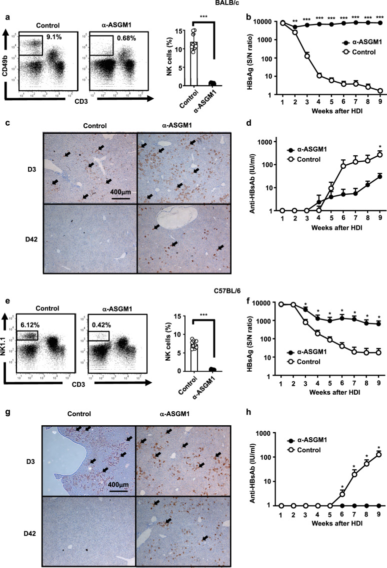
Fig. 1.
Anti-asialo GM1 (ASGM1) antibody treatment significantly impaired hepatitis B virus (HBV) clearance. a BALB/c mice at 6–7 weeks old were treated with anti-ASGM1 antiserum or an isotype control. Intrahepatic leukocytes were isolated, and the natural killer (NK) cell frequency was analyzed by flow cytometry. A representative flow cytometric analysis of NK cells (left) and their frequency (right panel) is shown. b–d BALB/c mice (n = 10) underwent hydrodynamic injection (HDI) with adeno-associated virus (pAAV)/HBV1.2 plasmids in the presence or absence of anti-ASGM1 treatment. Anti-ASGM1 treatment was performed twice per week over the detection period. Serum titers of HBsAg (b) and anti-HBs Ab (d) at the indicated times were determined by ELISA. c IHC staining for HBcAg expression in the livers (arrow) of anti-ASGM1-treated BALB/c mice compared to that in the livers of control mice on days 3 and 42 after pAAV/HBV1.2 injection. Data are shown as the mean ± SEM. *p < 0.05, **p < 0.01, ***p < 0.001 between selected relevant comparisons by Student’s t-test. e C57BL/6 mice (n = 10) at 6–7 weeks old were treated with anti-ASGM1 antiserum or an isotype control. Intrahepatic leukocytes were isolated, and the natural killer (NK) cell frequency was analyzed by flow cytometry. A representative flow cytometric analysis of NK cells (left) and their frequency (right panel) is shown. f–h C57BL/6 mice (n = 10) were HDI with pAAV/HBV1.2 plasmids in the presence or absence of anti-ASGM1 treatment. Anti-ASGM1 or isotype antibody treatment was performed twice per week over the detection period. Serum titers of HBsAg (f) and anti-HBsAb (h) were measured at the indicated times. g IHC staining of HBcAg expression in the liver (arrow) on days 3 and 42 after pAAV/HBV1.2 injection. Data are shown as the mean ± SEM. *p < 0.05, **p < 0.01, ***p < 0.001 between selected relevant comparisons by Student’s t-test

