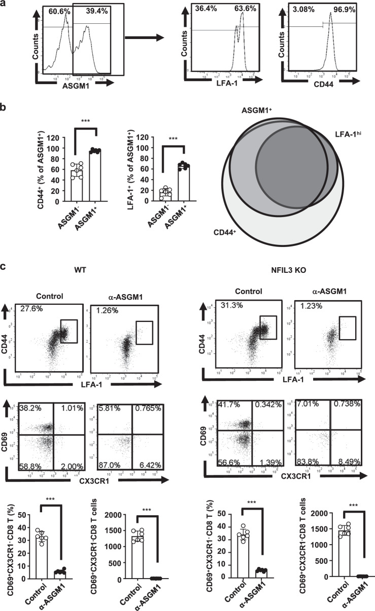
Fig. 5.
The major intrahepatic ASGM1-positive immune cells in NFIL3 KO mice were CD44+ LFA-1+ CD8 T cells. a Intrahepatic leukocytes (IHLs) from naive NFIL3 KO mice were isolated. Among CD8 T cells, the expression of ASGM1, CD44, and LFA-1 was examined. b Quantification of CD44+ and LFA-1+ cells among ASGM1+ CD8 T cells from (a) is shown. The right figure presents a Venn diagram showing the overlap among ASGM1-, CD44-, and LFA-1-positive cells from each group. c C57BL/6 (n = 6) and NFIL3 KO mice (n = 6) were injected with 20 μl of an anti-ASGM1 Ab. One day later, IHLs were harvested, stained, and analyzed by flow cytometry. A representative flow cytometric analysis of CD44+ LFAhi and CD44+ CD69+ CX3CR1− CD8 T cells in both wild-type and NFIL3 KO mice and their frequency, as well as cell numbers, is shown. Most of the CD44+ LFAhi, as well as CD44+ CD69+ CX3CR1− CD8 T cells in both wild-type and NFIL3 KO mice, were depleted after treatment with the anti-ASGM1 Ab. *p < 0.05, **p < 0.01, ***p < 0.001 between selected relevant comparisons by Student’s t-test

