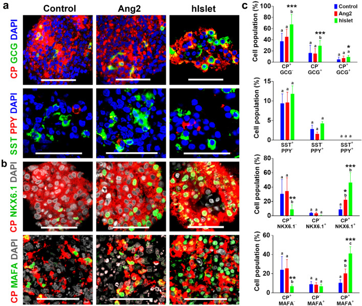
Figure 2.
Representative organogenesis of iPSC-derived islet organoids. At the end of differentiation, the islets were immunofluorescently labeled for (a) C-peptide (CP, red) and glucagon (GCG, green), somatostatin (SST, green) and pancreatic polypeptide (PPY, red). (b) NKX6.1 (green) and CP (red), and MAFA (green) and CP (red). Cells were counterstained with DAPI (grey). Scale bars, 50 μm. Human islets (hIslet) served as a positive control. (c) Semi-quantitative analysis of cellularity of the islets. Image analysis was performed using ImageJ software (n = 7–16 images for each condition). Results are shown as mean ± SD. Different letters indicate significant differences between the groups and p-value represented as *p < 0.05; **p < 0.01; and ***p < 0.001 compared to the control group.

