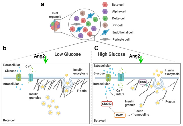
Figure 8.
Illustration of Ang2 effect on iPSC islet differentiation and iPSC-derived islet microenvironment. (a) Ang2 supports islet differentiation with the generation of β, α, δ, PP-cells and additionally, promotes the generation of endothelial and pericyte cells. (b-c) iPSC-derived islets under Ang2 cue showed Ca2+ influx and regulated activity of CDC42-RAC1 pathway synchronous with glucose level change, leading to F-actin remodeling aided by gelsolin for physiologically functional insulin exocytosis. Created with BioRender.com.

